

The causes of tooth decay—commonly referred to as “cavities”—have long been known: defects in the tooth’s outer layer, excessive carbohydrate intake, and inadequate oral hygiene.
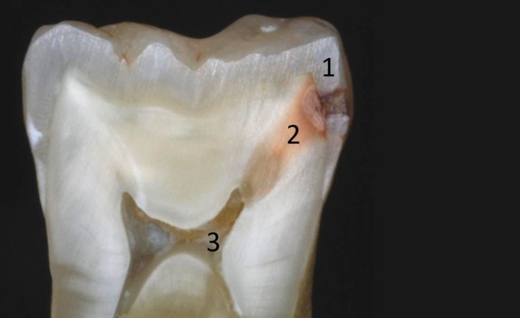
After a certain amount of mineral content has been lost from the tooth enamel, bacteria penetrate the tooth’s protective outer layer (1) and, by destroying the dentin (2), reach the pulp chamber (3) inside the tooth where the nerve is located. This process can take years to unfold, but it may also occur within just a few months.
It is the dentist’s responsibility to carefully examine the visible tooth surfaces of patients who attend check-ups—even when they have no symptoms—and to detect the presence of any of the above processes.
If the patient presents in time (at stages 1 or 2) and the destruction of the tooth crown is not yet extensive, further damage can be halted with the least invasive dental treatment. The tooth can be restored to its original shape, regaining full function, easy cleanability, and aesthetics—without additional complications. Achieving this at the highest professional standard and with maximum precision is possible using a dental microscope capable of up to 25× magnification.
Microscope-assisted fillings offer numerous advantages over conventional fillings. The most important benefits include:
1. Greater precision
Using a dental microscope with up to 25× magnification, the dentist can view the tooth in great detail, allowing for much more precise removal of decayed areas and more accurate placement of the filling.
2. Less tissue loss
Thanks to the enhanced visibility, only the truly affected areas are removed, allowing the healthy tooth structure to be preserved.
3. Longer-lasting filling
Because of the precise fit, there is a lower risk of the filling separating from the tooth or developing decay underneath, which can significantly extend the filling’s lifespan.
4. Better aesthetic result
Thanks to the meticulous work and the use of high-quality filling materials, the fillings achieve a more natural and visually pleasing appearance.
5. Lower risk of future problems
Using a microscope makes it easier to detect hidden cracks, decay, or other issues that are not always visible to the naked eye.
6. Better hygiene and reduced risk of infection
During microscope-assisted treatment, there is a much lower chance of leaving behind infected or decayed tissue, which reduces the risk of future inflammation or reinfection.