
The condition of the teeth has traditionally been assessed— and is still assessed today— by means of an oral examination supplemented with X-ray imaging.
With the advent of magnification, the examination of teeth has undergone remarkable development. It makes a significant difference whether the teeth are inspected with the naked eye or under 25× magnification. Detecting early signs of decay or cracks caused by overloading in good time can prevent the need for root canal treatment and, ultimately, the loss of the tooth.
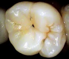
Will the dental treatment stop at a small, barely noticeable filling, or—because of the excessive progression of decay—will a root canal eventually be required?
If root canal therapy has already become unavoidable, is it still worthwhile to save the tooth? Is there enough sound tooth structure left to allow for a successful restoration after the root canal?
All of this makes it clear that the factor of time is of outstanding importance.
When a patient comes in for an oral examination, it is our responsibility as dentists to ensure that no hidden changes—even those causing no symptoms—remain undetected.
Microscopic examination—performed under high magnification—is the most thorough method for patients who value detailed knowledge of their dental health. It provides reliable information about the condition of the teeth and ensures timely awareness of any necessary or urgent treatments.
Microscopic diagnostics always go hand in hand with photographic documentation: high-resolution images are taken of every finding. This not only supports the diagnostic process, but also makes it easier to explain the problem to the patient and, not least, helps build an atmosphere of trust between patient and dentist